6901 Helen of Troy, Suite E2, El Paso, TX 79911
(915) 581-3391

Dental CBCT Scan in El Paso

Westside Endodontic Associates

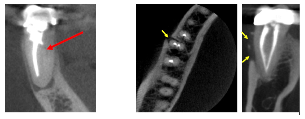
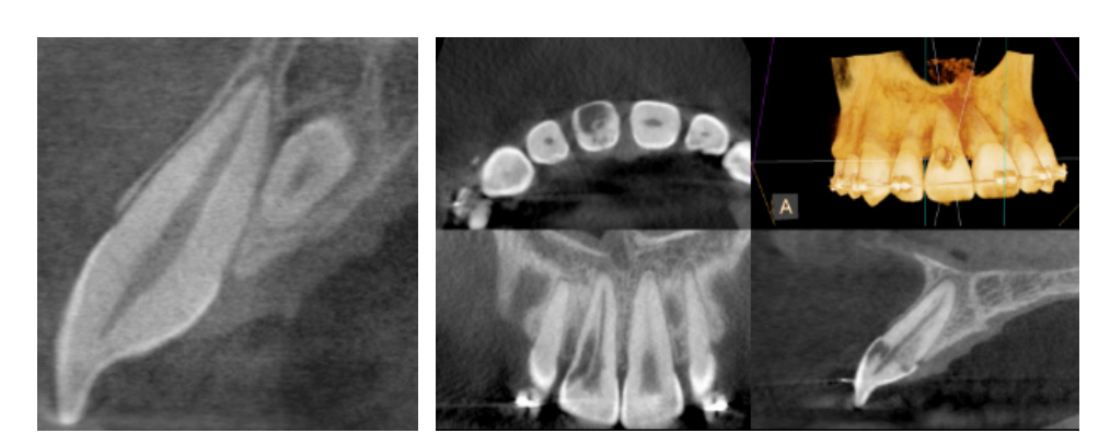
Cone beam computed tomography (CBCT) offers accurate and high quality 3-D representations of dental and facial structures. This enables our endodontists to more precisely diagnose an underlying problem, detect unforeseen defects, and minimize the need for exploratory procedures. CBCT facilitates more effective care and more consistent successful outcomes.

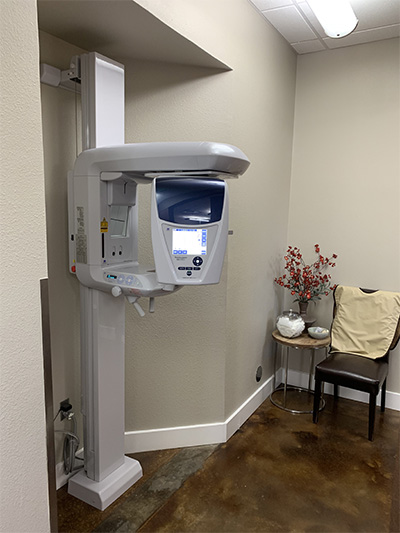
At our office we utilize the J. Morita Veraviewepocs 3D F40, which offers the highest resolution 3-D imaging at the lowest radiation dose.

Clinical applications of CBCT technology in endodontics include the following:

  • Detection and diagnosis of pathology at the root end of the teeth
  • Pre-operative assessment of the dental surgical area and treatment planning
  • Assessment and management of traumatic dental injuries
  • Evaluation of root canal anatomy and complex morphology
  • Detection of root fractures and root resorption
  • Evaluation of previously endodontically treated teeth
  • Dental implant work-up

Occasionally, dental defects or pathology that deem a tooth non-restorable will be revealed on a CBCT scan. If this happens, the endodontist will advise you that the tooth cannot be saved.

(Pictures of missed canal, vertical root fracture, extra tooth, and resorption)

Westside Endodontic Associates

Westside Endodontic Associates

Here are some ways the cone beam CT has helped us to be better clinicians and provide the best treatment possible:

This patient presented with pain on her upper right molar. The first x-ray is a periapical film, which shows us that a root canal procedure has already been completed on tooth #2. The periapical film also shows infection present around the mesio-buccal root, but it does not provide much information as to why there is infection. The cone beam CT give us much more information. As seen here, it shows that a canal was missed when the original RCT was completed. For treatment purposes it gives us a good road map of where to go to find this canal so that it can be properly cleaned and the patient’s infection can heal.

Westside Endodontic Associates

On this patient’s periapical x-ray we see evidence of resorption (invasive resorption). The cone beam image was very helpful in determining if this tooth could even be saved. Sometimes the resorption is so extensive that the tooth will need to be extracted. In this case, the cone beam CT helped us make the decision to try and save the tooth through root canal treatment.

This patient presented with pain on her upper right side. The periapical x ray showed that root canal treatment had already been performed on tooth #2. The treatment looked well done, and it was a little confusing as to why the patient was in pain. After taking a Cone Beam CT, we discovered that the root filling material in the mesio-buccal canal was extruded out of the root end and into the buccal tissue. This was the source of the patient’s pain. This finding on the Cone Beam CT allowed us properly diagnose and formulate a treatment plan to alleviate the patient’s pain.

Westside Endodontic Associates

Although we have a good understanding of the root canal anatomy in most teeth, there are still teeth that have a complex anatomy that only Cone Beam CT imaging can bring to our attention. Below is a case where the cone beam CT showed us a very unusual anatomy and helped us treat the tooth successfully.

Westside Endodontic Associates

This patient was having pain on the lower left side. No infection or problems can be seen on the periapical x ray. However, on the cone beam CT, we see clear evidence that the root is cracked. This information is important, because retreating this root canal would have a hopeless prognosis due to the crack. Instead the patient was referred for extraction and implant. Time and money were saved and the correct treatment plan was selected.

Westside Endodontic Associates